An echocardiogram uses ultrasound waves to produce images of
your heart. This common test allows your doctor to see your heart beating and pumping blood. Your doctor may suggest an echo for problems with the valves or chambers of your heart.
A Contrast Echocardiogram is a specific type of echocardiogram which uses a special ultrasound enhancing agent that is injected into one of the veins in your arm to help show structures in the heart better. It allows the inside of the heart to be seen more clearly on the ultrasound pictures.
Additional note for Contrast Echo:
There is an extremely small risk (less than 1 in 10,000) of developing an allergic reaction to the agent used. Uncommon but possible side effects can include dizziness, weakness,fatigue, palpitations, headaches, and nausea.
Let the technologist know if you have any previous allergic reaction to echo contrast (called perflutren), blood, blood products, albumin, if you are pregnant or breastfeeding, if you have had any recent procedures.
Test Process
The test will take about 45 minutes.
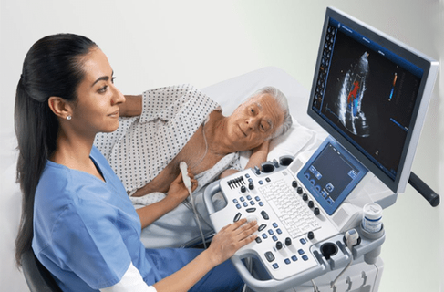
You will be asked to remove your shirt and put on a gown to keep you comfortable and maintain privacy. While lying on an examination table, a technician will apply a colorless gel to your chest and then a transducer will be moved back and forth across different areas to obtain multiple views of your heart. You may be asked to move from your back and to the side. Instructions may also be given for you to breathe slowly or to hold your breath. This helps in obtaining higher quality pictures.
With Doppler echocardiograms, as the transducer moves over your heart, you will hear a “whooshing” sound, much like that of a washing machine. This sound relates to the movement of blood within your heart chambers. The images are constantly viewed on the monitor and recorded for a permanent record of the examination. This is reviewed by the physician prior to completion of the final report.